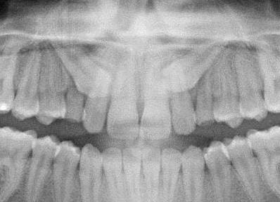
Is 3D imaging useful for diagnosing impacted canines?
It is essential that we accurately identify the position of impacted maxillary canines. In the past we have relied on using 2D views. Nevertheless, with the advent of CBCT imaging, it is now possible to obtain detailed 3D images. However, can we identify the severity of the impaction, and does this change our treatment decisions using 3D views? This interesting new paper answers these question very effectively.
I discussed research into a similar question in 2017. We concluded that CBCT is more accurate than 2D imaging techniques. However, no robust evidence supported CBCT as the first-line impacted canine imaging technique. This controversial paper generated a great deal of discussion on my blog. This new study is, therefore, timely

A multi-national team did this study. The AJO-DDO published the paper.
Three-dimensional decision support system for treatment of canine impaction
Dylan J. Keener et al.
AJO-DDO Advance access: DOI: https://doi.org/10.1016/j.ajodo.2023.02.016
What did they ask?
They did this study to:
“Develop a 3D characterisation of the severity of maxillary impacted canines and test the clinical performance of this as a treatment decision support tool”.
What did they do?
The study team did an extensive study and analysed CBCTs taken to diagnose patients with maxillary-impacted canines.
They included images of patients over 12 years old with either unilateral or bilateral impacted canines. The investigators obtained these records from their clinical databases.
They then obtained the following information from the CBCTs.
- The amount of root resorption of the maxillary central and lateral incisors.
- Data on the amount of height, pitch and roll of the canines.
- The amount of sagittal overlap of the lateral incisor.
They combined the information on the height, pitch, roll and the amount of overlap to calculate a severity index. The higher the score represented a more challenging clinical scenario. They then used this data to construct a 3D clinical report.
In the next stage of the study they looked at the clinical effectiveness of the report in decision-making. They followed these stages:
- Six part-time orthodontic faculty members at the University were enrolled in the study.
- The study team selected ten sets of patient records from the files at the University of Michigan. These included three patients with bilateral and seven with unilateral impactions. Six of these were palatal, four bicortical and three buccal.
- They asked the orthodontists to look at the patients’ photographs, panoramic and cephalograms (traced and untraced measurements).
- The orthodontists completed a short questionnaire that collected information on their perception of the canine and treatment decisions.
- Two weeks later, they viewed the relevant 3D diagnostic reports, repeated their decisions, and recorded how the CBCT report affected their decision.
What did they find?
Firstly, they found that the method of constructing the severity index was very reproducible.
When they looked at the data on the canines they found:
- The most significant contribution to the index was overlap.
- Palatally placed canines showed the most significant contribution to the overlap score.
- Canines in a bicortical position had the highest severity rating for resorption.
When they looked at the treatment decisions;
- The 3D report changed most of the decisions for categories of choice of mechanics, patient/parent education and estimated treatment time.
- Overall 22% of decisions were changed about exposure and traction vs extraction.
Their conclusions were:
“It was possible to construct a severity measure for impacted canines”.
“Clinicians preferred CBCT to conventional imaging methods”.
‘The 3D report had a profound impact on the clinicians’ treatment decisions”.
What did I think?
This was a very complex study to do. The team must have spent a significant amount of time and effort in completing it. This is a very well-written paper that substantially contributed to our knowledge.
When I interpreted the paper, the research team provided information on the limitations of their study. Importantly, it was cross-sectional. Ideally, the treatment outcomes should have been included in the validation data. Significantly, the strength of the evidence would have been increased if they had included more clinicians in the treatment decision exercise. It was good to see that they had listed this information in their paper instead of brushing it under the carpet.
They also provided a way forward for future research. This included testing the index over many sites before implementation. Importantly, they highlighted using machine learning to produce detailed diagnostic input for clinicians. This may be built upon by using AI to make treatment decisions in a similar way to the paper that I recently discussed on extraction decisions. We are heading towards a brave new world of automated orthodontic treatment decisions!

Emeritus Professor of Orthodontics, University of Manchester, UK.
Thanks for sharing dear professor, so do you it’s worth requesting a cbct whenever we encounter an impacted canine?
Thanks alot dear professor.
My question about the the possibility of the ankylosis of impacted canines ?
I think it’s the most challenging issues in treating the impacted teeth and also main issue related to the prognosis! They didn’t mentioned
Regarding treatment decision with and without the use of 3D imaging there is a well-written paper with high evidence from 2006.
Bjerklin K, Ericson S. How a computerized (CT) examination changed the treatment plans of 80 children with retained and ectopically positioned maxillary canines. Angle Orthod 2006;76:43-51.
Thanks for the analysis of the paper
My question is when should CBCT be used for ectopic canines?
I have some specialists wanting them for every case and in view of the increased radiation dose I am not sure whether they are indicated
Of course some are if potential or actual resorption is suspected
Are there guidelines?