A new review of adenoid, tonsil and craniofacial growth.
Over the past few years, there has been growing emphasis on the relationship between craniofacial growth, airway dimensions, and obstructive sleep apnoea in children. As a result, enthusiasm is growing for orthodontic interventions aimed at improving children’s breathing. Importantly, the evidence underpinning these treatments is somewhat lacking.
This new paper by Audrey Yoon and her colleagues addresses an important aspect of the debate underpinning this treatment. The paper examines the growth of the adenoids, tonsils, and tongue in relation to craniofacial development. Importantly, it raises questions about how we interpret anatomical findings and how they may influence orthodontic treatment.

Conceptual growth curves of oropharyngeal soft tissues and craniofacial skeletal structures: clinical implications for paediatric airway management
Audrey Yoon et al
Sleep and Breathing Advance access. https://link.springer.com/article/10.1007/s11325-026-03809-6
What did they ask?
They did this review to
“Develop an updated understanding of the growth of the adenoids, palatine tonsils, lingual tonsils, and tongue in relationship to the developing craniofacial skeleton”
“Develop an updated understanding of the growth of the adenoids, palatine tonsils, lingual tonsils, and tongue in relationship to the developing craniofacial skeleton”
What did they do?
To answer this question, they conducted a narrative review. We need to bear in mind that this was not a systematic review or clinical trial; as a result, it is very low-level evidence. I will expand on this later.
What did they find?
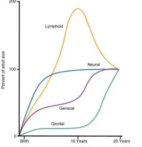
They began their assessment by noting that the classic Scammon’s growth curves, which we have used for years, are not necessarily accurate. One key point was that the classic curves show lymphoid tissue size increasing to about 200% of adult size around puberty, followed by a period of regression. Importantly, they noted that the original lymphoid curve was derived largely from thymus measurements and may therefore be less relevant to contemporary orthodontics.
They then continued to review the literature on the various tissues. This was a highly detailed review, and I don’t have space to cover every point in this blog. However, I felt that these were the main points they raised.
Adenoids
The adenoids reach their maximum size earlier than we thought. The authors felt that this occurred between 6 and 8 years of age. They then remain relatively stable before gradually reducing in size.
Palatine tonsils
The palatine tonsils follow a distinct growth pattern, undergoing rapid growth in early childhood and then gradually decreasing in size. The evidence was not entirely consistent. For example, some ultrasound studies showed that tonsillar volume increases during the first 3 years and then changes relatively little until approximately 12 years old. This also illustrates that different measurements reveal different anatomical characteristics.
Tongue
They also examined tongue growth, and their review suggested that the anterior two-thirds of the tongue approaches adult dimensions between 8 and 10 years, whereas the posterior third continues to grow into adolescence.
In summary, airway dimensions appear to be influenced by both changes in tonsil size and craniofacial growth. This is not new. However, the team suggests that the timing of this differs from what we have traditionally accepted.
The window of vulnerability
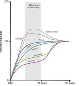
They then revisited the concept of the “window of vulnerability”, as described in a previous publication. This window occurs between 5 and 10 years of age. During this period, the adenoids and tonsils are relatively large, while the surrounding craniofacial structures are still developing. As a result, lymphoid tissue may occupy a greater proportion of the available airway space. However, as the child grows, the craniofacial skeleton and pharyngeal airways enlarge, reducing the relative contribution of lymphoid tissue to airway narrowing.
This leads me to consider that many studies of treatments during this period may be reporting on simple natural growth. We need to bear this in mind when we interpret any of this work.
Does the window of vulnerability also apply to “selling” unnecessary treatment to vulnerable families? Perhaps we can discuss this?
What did I think?
This was an interesting paper, but we need to remember that this was a narrative review. Unfortunately, this form of review does not provide the same level of protection against selection bias as a systematic review. Importantly, there was no formal assessment of the certainty of the evidence. This was evident because several key publications they cited were also narrative reviews. Furthermore, many of the studies included in the review used a wide range of measurements and methods. This means there must be marked heterogeneity within the review that is neither measured nor stated. The quality of evidence is very low.
I was also unsure how they generated the data in their “window-of-vulnerability” figure. To examine this further, I looked closely at the adenoids section. It appears the authors drew their conclusions from 10 publications, mostly retrospective or cross-sectional, using various measurements across mixed age groups. When I reviewed the data in these papers, I could not see it reflected in the figure.
The authors address this in the text, noting that this figure is a conceptual schematic illustration. This is an important distinction because the figure gives the impression that the authors have precise data describing the growth of the adenoids, palatine tonsils, tongue, maxilla and mandible from birth to adulthood. However, the paper does not present a new longitudinal dataset from which these curves were calculated. Importantly, there is little evidence to underpin this figure and we should be cautious.
I also noted that the authors concluded that we need to recognise the window of vulnerability, which is important for early assessment and timely intervention. While I agree that children with symptoms of obstructive sleep apnoea should be appropriately assessed, this paper’s results do not establish that orthodontic treatment during this window is effective or necessary. Interestingly, the authors made no clear recommendations on this.
Importantly, we should not assume that identifying a narrow airway means that orthodontic treatment is necessary. We also should not suggest that expanding the maxilla will prevent obstructive sleep apnoea because it changes nasal cavity dimensions. These claims require evidence from clinical trials, which are currently lacking.
The biggest takeaway from this paper was that if airway dimensions and lymphoid tissue proportions change naturally throughout childhood, we must be very careful about attributing improvements in breathing or airway size to orthodontic treatment. As a result, I feel the review strengthens the argument against drawing causal conclusions from uncontrolled studies of orthodontic care. Natural growth changes the relationship between lymphoid tissue and airway size, which can produce improvements that may be mistakenly attributed to treatment. Perhaps watchful waiting is the way forward for many children? But this is controversial.

Emeritus Professor of Orthodontics, University of Manchester, UK.
Dear Prof. O´Brien,
Thank you for bringing up the paper by Yoon et al.
As you wrote they had made “a highly detailed review” on growth of various soft tissues possibly having an impact on airway dimensions. To me their narrative review is quite fine provided that they do not recommend early ortho treatment based on the findings.
But I was suprised to read their comments on Scammon’s growth curves with a new figure. This point has been my in my lectures for the past 20 years. And more importantly published in the Mosby´s Orthodontic Review, 3rd Ed. in July 2025, Chapter 5 “Development of Ortofacial Function” by Gregory Antonarakis and Stavros Kiliaridis and me.
Let me know if you don´t have access to this book. If needed I can send you the chapter with the figure.
Thanks for the excellent Blog!
Well done Kevin!
I agree with you, Kevin:
Apart from the subtle confirmational bias, some significant omissions are evident, including those noted by Dr Peltomaki. For clinical considerations, the entire ecosystem of upper airway behavior needs to be addressed [1] instead of a piecemeal, reductional approach.
1. Singh GD, Battle J. Upper airway homeostasis: A review. Sleep Med. 2026;148:109221.