Mini screw anchored maxillary protraction for maxillary deficiency works!
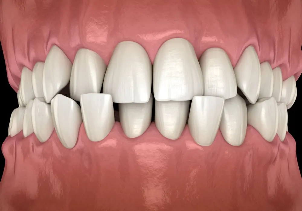
The correction of Class III malocclusion is one of the most challenging orthodontic treatments. Over many years clinicians have been searching for effective methods of changing skeletal growth for this group of patients. One of these has been Bone Anchored Maxillary Protraction. This method has attracted much interest, despite no randomised trials into this intervention.
Recently this technique has been modified with the development of Miniscrew Assisted Maxillary Protraction (MAMP). This method uses mini screws in the mandible and a miniscrew anchored Hyrax appliance in the maxilla instead of mini plates.
This paper outlines the first randomised controlled trial of this relatively new technique.
A team from Egypt did this study, and Progress in Orthodontics published the report.
Ahmed Mohamed Kamel et al.
Progress in Orthodontics: Advanced Access DOI: https://doi.org/10.1186/s40510-023-00473-4
One good thing about this journal is that it is open-access for all papers. As a result, you can easily have a look at the report.
What did they ask?
The team wanted to:
“Evaluate the effects of miniscrew-anchored maxillary protraction (MAMP) and compare them with the growth changes in an untreated control group in growing patients with Class III malocclusion”.
What did they do?
The investigators conducted a two-armed parallel-group randomised controlled trial with a 1:1 allocation.
The PICO was:
Participants:
Forty patients with skeletal Class III malocclusion (ANB <0, Wits <-2). They needed to be growing with CVM stages of CS1-CS3 in the late mixed or early permanent dentition.
Intervention
Maxillary protraction with Class III elastics anchored by Hybrid Hyrax to miniscrews in the maxilla and a bar attached to two miniscrews in the mandible between the canine and lateral incisor.
Control
Untreated participants that had similar inclusion criteria.
Outcomes
Maxillary position according to change in A Point. Secondary outcomes were many cephalometric changes.
The team recorded the data before and after the maxilary protraction period of treatment. They defined this as when the participant had a 2-3mm positive overjet. The second data collection stage for the control group was 12 months after enrolment.
The statisticians did a sample size calculation based on previous studies. This work revealed that they needed to enroll 20 patients to achieve a target sample size of 14 participants per group.
They used pre-prepared block randomisation. A person who was not involved in the trial’s day-to-day running kept this list. They did not provide further data on allocation concealment.
They did the relevant statistical analysis using an Intention to Treat method that enables them to impute data for any missing values.
What did they find?
The team enrolled 40 participants in the trial, 20 per group. Three of the MAMP group dropped out (1 moved away, and two discontinued treatment). However, seven in the untreated control group dropped out (3 were lost, and four refused to continue in the trial). The team reported that they analysed 17 MAMP and 13 control patients.
Importantly, the groups were balanced for gender and age. The mean length of protraction treatment was 11.9 months. The second data collection stage for the control group was 12 months.
They presented data on a complete case and ITT basis. I will only mention the ITT data as this includes adjustments for post-treatment measurements with covariates of pre-treatment values.
They produced a large amount of data across many cephalometric measurements. Aside from making it difficult to read, this method also risks false positives and “statistical fishing”. As a result, I concentrated on the primary outcome measure of the maxillary position, skeletal relationship, and incisal angulation. I decided to look closely at the final measurements as this gives information on the final outcomes. I prepared this data in this table.
| Variable | Control | Treatment | Difference | 95% CI | p |
| A-VR (mm) | 57.8 | 60.5 | 2.99 | 1.6-4.5 | 0.001 |
| SNA (0) | 78.3 | 81.5 | 3.24 | 1.2-5.15 | 0.001 |
| Wits (mm) | -4.0 | -0.07 | 3.95 | 1.88-6.01 | 0.001 |
| Is-PP (0) | 116.35 | 117.35 | 1.18 | -0.3-7.7 | 0.132 |
| Ii-MP (0) | 87.2 | 87.6 | 0.42 | -0.22-1.0 | 0.74 |
Their conclusion was:
‘The MAMP treatment effectively increases forward growth of the maxilla”.
However, they drew attention to the high attrition rate, and I will discuss this later.
What did I think?
This small study was interesting. The study team attempted a complex investigation to answer a clinically relevant question. I want to congratulate them on their work.
Unfortunately, they had a high dropout rate from their initially small sample. This means there is likely to be a marked level of uncertainty in their findings. I acknowledge that they took steps to impute missing data and conduct an ITT analysis. Nevertheless, this does not avoid problems with the low sample size. This is evident from most of the data’s wide 95% confidence intervals.
As a result, we must consider that this is a good pilot study. In this respect, the study has some value.
Bearing this in mind, we can conclude that MAMP may have the potential for the treatment of developing Class III problems. It certainly, is less invasive than placing bone anchors. Does this data provide us with sufficient evidence to adopt this technique? It is, undoubtedly, better than the data used to support BAMP.
However, It is up to you as clinician-scientists to take this decision with your patients. This study provides good data for sample size calculations for more extensive investigations into this treatment. I hope that the current team can take this type of study forwards.

Emeritus Professor of Orthodontics, University of Manchester, UK.
According to the literature a much simpler RPE + FM in mixed dentition has about 70% chances of success.
The 30% of the remaining patients grow as untreated Class 3 with longer growth spurt, longer mandible (3 times more than controls in males) up to CVM stage 5-6 …..
For how long should they continue to wear elastics maintaining Tads? From late mixed dentition to 17 yrs.old ???
Is there any study demonstrating that the Liou Tq. makes the forward movement of the maxilla in mixed dentition bigger ? As far as I know only 1 study by Costanza Miazzini on cleft patients.
The criteria of success is only occlusal.
Many patients at age 17 elects to go for surgery to improve their appearance other than the overjet.
This presentation shows some promise for the future tx of young class III patients but my first thoughts are about the mandibular TADs and failure rate. One of the authors (Wilmes) has stated in a recent lecture that he does not like to insert TADs in the mandible younger than age 14. My personal experience seems to support that kind of high failure rate from lower jaw screws.
I realize the idea of TADs saves the need of surgery to do Bollard plates or a mentoplate but I am worried about failure. The same author referred to above has stated that his group has a 100% success rate with the mentoplate at least regarding failure. I am skeptical that that kind of success can be duplicated with TADs.
Comments?
The numerical values are interesting, Would have been nice to see some pre and post cephs to visually see the treatment effects.